

INTRODUCTION
Microbiology is the study of organisms too small to be seen distinctly with the unaided eyes. The nature of this discipline makes the microscope of crucial importance because the study of microorganisms is impossible without the microscope. Microscopes provide magnification which enables us to see microorganisms and study their structures. The magnification attained by microscopes range from x100 to x400,000 in addition there are different types of microscopes and many techniques have been developed by which specimens of microorganisms can be prepared for examination. This unit examines the different types of microscopes, how the microscopes work and how specimens are prepared for examination.The Microscope
A microscope is an instrument for producing enlarged images of objects too small to be seen unaided.
Types of Microscopes
Microscopes are of two types:- Light (optical) and
- electron depending on the principle on which magnification is done.
The Light Microscope
This is a type of microscope in which magnification is obtained by a system of optical lenses using light waves. It includes:- Bright Field Microscope
- Dark Field Microscope
- Fluorescence Microscope
- Phase Contract Microscope
The Bright Field Microscope
- The ordinary microscope is called a bright field microscope because it forms a dark image against a brighter background.
- The microscope consists of a sturdy metal body or stand made up of a base and an arm to which the remaining parts are attached.
- A light source, either a mirror or an electric illuminator, is located at the base.
- Two focusing knobs, the fine and coarse adjustment knobs are located on the arm and can move either the stage or the nose piece to focus the image.
- The stage is positioned about halfway up the arm and hold microscope slides by slide clips or a mechanical stage clip.
- There is a substage condenser mounted within or beneath the stage which focuses a core of light on the slide.
- The upper part of arm of the microscope holds the body assembly to which a nose piece and one or more eyepieces or ocular lenses are attached.
- Most advanced microscopes have eyepieces for both eyes and are called binocular microscopes.
- The nose piece holds three to five objective lenses of different magnifying power and is easily rotated to position any objective.
- The image you see when viewing a specimen is focused by the objective and ocular lenses working together.
- Light from the specimen which has been illuminated is focused by the objective lens creating an enlarged image within the microscope. The ocular lens further magnifies this primary image.
- The total magnification is calculated by multiplying the objective and eye piece magnification together; e.g. if a 45x objective is used with a 10x eyepiece, the overall magnification of the specimen will be 450x.
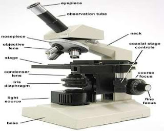
Fig. 1: The Bright Field Microscope
Source: http://biology.unm.edu/ccouncil/Biology_203/Images/
Microscopes/microscope6.jpeg
The Dark-Field Microscope
The dark field microscope is used to observe living unstained cells and organisms as a result of change in the way they are illuminated.A hollow core of light is focused on the specimen in such a way that unreflected and unrefracted rays do not enter the objective only light that has been reflected or refracted by the image forms an image.
The field surrounding the specimen appears black while the object itself is brightly illuminated.
The dark field microscope is useful in revealing many internal structures in larger eukaryotic microorganisms. It is also used in the examination of unstained microorganisms suspended in fluids, e.g. wet mount and hanging drop preparation.
The Phase-Contrast Microscope
This type of microscope converts slight differences in refractive index and cell density into easily detected variations in light intensity and is used to view living cells. The background formed by the undeviated light is bright while the unstained objects appear dark and well defined. This microscope is very useful for studying microbial motility, determining the shape of living cells and detecting some bacterial components such as endospores and inclusion bodies. It is also used in studying eukaryotes.The Fluorescent Microscope
This type of microscope exposes a specimen to ultraviolet, violet or blue light and forms an image of the object with resulting fluorescent light. The most commonly used fluorescence microscope light is epifluorescence microscope which is also called incident light or reflected light microscope. Epifluoresence microscope employs an objective lens that also acts as a condenser. A mercury vapor arc lamp or other source produces an intense beam of light that passes through an exciter filler. The exciter filler transmits on the desired wavelength of excitation lightThe excitation light is directed down the microscope by a speed minor called the dichromatic minor. This minor reflects light of shorter wavelength but allows light of longer wavelength to pass through. The excitation light continues down through the objective lens to specimen stained with spaced dye molecules called fluorochromes.
Microscope Resolution
Resolution is the ability of a lens to separate or distinguish between small objects that are close together, i.e. the microscope must produce a clear image and not just a magnified one. It is also known as the resolving power. Resolution is described mathematically by an equation in the 1870s by Ernest Abbe, a German physicist. The Abbe equation states that the minimal distance (d) between two objects that reveal them as separate entities depends on the wavelength of light (l) used to illuminate the specimen and on the numerical aperture of the lens (nsinq) which is the ability of the lens to gather light.The wet mount or hanging drop technique
The technique permits examination of organisms in a normal living condition. A wet mount is made by placing a drop of fluid containing the organisms on a glass slide and covering the drop with a cover slip. Petroleum jelly may be used to provide a seal between the slide and covers slip after which the slide is viewed under the microscope.This method is desirable because,
- it prevents distortion of the morphology of spiral bacteria when they are stained and dried.
- it reveals whether organisms are motile or not.
- some cell inclusion bodies are easily observed.
- spore formation and germination may also be observed in living cells.
Fixed, Stained Smears of Microorganisms
These are frequently used for the observation of the morphological characteristics of bacteria. The procedure makes the cell more clearly visible, and differences between cells of different species and within the same species can be demonstrated. The essential steps in this procedure are:- preparation of the film or smear
- fixation and
- application of one or more staining solution.
Fixation
Fixation is the process by which the internal and external structures of cells and microorganisms are preserved and fixed in position. It in-activates enzymes that might disrupt cell morphology and tough cell structures so that they do not change during staining and observation. A microorganism usually is killed and attached firmly to the microscope slide during fixation.There are two fundamentally different types of fixation.
- Heat Fixation: Is routinely used to observe prokaryotes. Typically, a film of cells (a smear) is gently heated as a slide is passed through a flame. Heat fixation preserves overall morphology but not structures within cells.
- Chemical Fixation: Is used to protect fine cellular sub-structure and the morphology of larger, more delicate micro organisms. Chemical fixatives penetrate cells and react with cellular components, usually proteins and lipids, to render them inactive, insoluble, and immobile. Common fixative mixtures contain such components as ethanol, acetic acid, mercuric chloride, formaldehyde, and glutaraldehyde.
Staining of Specimens
Although living microorganisms can be directly examined with the light microscope, they often must be fixed and stained to increase visibility, accentuate specific morphological features, and preserve them for future study.Types of Staining
Simple staining
This is a kind of staining in which a single stain or dye is used. Basic dyes such as crystal violet, methylene blue, and carbolfuchsin are used in simple staining to determine the size,shape and arrangement of prokaryotic acids.
Differential staining
These are staining procedures that make visible the differences between bacterial cells or part of a bacterial cell. It usually involves more than one dye used for staining.Gram staining
The Gram stain was developed in 1884 by the Danish physician Christian Gram. It is the most widely used differential staining procedure.SUMMARY
In light microscope, magnification is obtained by a system of optical lenses using light waves. Many types of light microscopes have been developed. They include bright fields, dark field, phase contrast and fluorescence microscope.- Electron microscope uses a beam of electron in place of light waves to produce the image of an object.
- The ordinary compound microscope is called the bright field microscope because if forms a dark image against a bright background.
- In the bright field microscope which is a compound the primary image is formed by an objective lens and enlarged by the eye piece or ocular lens to form the final image.
- The dark field microscope uses only refracted light to form an image and objects glow against a black background.
- The dark field microscope is useful in revealing many internal structures in larger eukaryotic microorganism.
- The phase-contrast microscope converts slight differences in refractive index and cell density into easily detected variations in light intensity and is used to view living cells, for studying microbial motility and detecting some bacteria components such as endspores.
- The fluorescent microscope exposes a specimen to ultraviolet, violet or blue light and forms an image of the object with resulting fluorescent light.
- Two general methods for preparing specimens for light microscope examination are the wet mount or the hanging drop technique and the dried fixed stained technique.
- A wet mount is made by placing a drop of fluid containing the organisms on a glass slide and covering the drop with a cover slip before viewing under the microscope.
- Fixation is a process by which the internal and external structures of cells and microorganisms are preserved and fixed in a position. It involves preparation of the smear, fixing with heat or chemical and application of one or more staining solutions.
- Electron microscopes use a beam of electrons to illuminate and create magnified images of specimens.
- Simple staining is a kind of staining in which a single stain or dye such as methylene and crystal violet is used. Differential staining involves the use of more than one stain or dye is used to make visible the differences between bacterial cells or part of a bacterial cell examples are the Gram stain.
Social Plugin