

INTRODUCTION
Bacteria are characterised based on the cell shape, size and structure cell arrangement, occurrence of special structures and developmental forms, staining reactions and motility and flagella arrangement. They are also characterised by the cell wall component, Gram stain reaction, cellular respiration and mode of nutrition. This unit examines the general characteristics of bacteria, shapes and forms of bacteria, structures external and internal in bacteria among other things.table of content
- composition of the microbial world
- general characteristics of bacteria
- historical aspects of microbiology
- a brief survey of microbes as friends and foes
- activities of living things
- cell activities
- fung and root
- general characteristics of algae
- general characteristics of fungi
- general characteristics of viruses
- general structure, anatomy, physiology of the root and the leaf
- microscope and specimen preparation
- prokaryotes and eukaryotes cells
- the relevance and scope of microbiology
- the cell, its general structure and activities
- the stem
- viruses-discovery/hiv-aids virus
General Characteristics of Microorganism
General characteristics of bacteria:
- They are prokaryotic
- They are simplest of all microbial cells
- Bacteria are single celled organisms
- They have distinctive cell wall which contain peptidoglycan
- They are measured in unit called micrometer
- Bacteria lack a true nucleus but have a region called the nucleroid region, i.e. DNA is free floating
- They may have additional DNA called a plasmid
- Their reproduction is by binary fission
- They are extremely diverse and numerous in soils and waters.
Size, Shape and Arrangement of Bacterial Cell
Size
Bacteria are very small, 0.5 to 1.0µm in diameter. Because of their small size, they have high surface area/volume ratio which results in a high growth and metabolism rate. No circulatory mechanism is needed for nutrients taken in because the mass of cell substance to be nourished is very close to the surface. Examinations of a microbial cell require the use of a high power microscope usually of about 1,000 diameters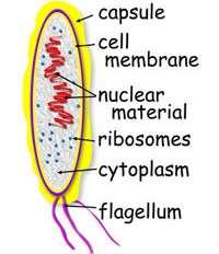
Fig. 1: A Basic Bacterium Cell
Source: Microorganisms in our World by Atlas (1995)
Shape and Arrangement
The shape of a bacterium is governed by its rigid cell wall which gives it a definite shape.Typical shapes of bacteria are:
- Cocci (Singular: Coccus), e.g. Staphylococcus
- Bacilli (rods) (Singular: rod, bacillus), e.g. Bacillus subtilis · Vibrios (Singular: Vibrio)
- Spirilla (Singular: Sprillum)
- Spirochaetes (Singular: Spirochaete), e.g. Treponema pallidum
- Cocci: They are round, oval or spherical in diameter characteristic arrangement when multiplying is based on arrangement of cells, they are called:
- Diplococci: cocci in pairs, e.g. meningococci and gonococci.
- Streptococci cocci in chains.
- Staphylococci: cocci in irregular clusters (like a bunch of grapes).
- Tetracocci: cocci in a group of four cells.
- Sarcinae: cocci in regular clusters.
Bacilli (Rod): These are stick like bacteria with rounded, square, tapered or swollen ends. They measure 1-10µm in length by 0.3- 1.0µm in width.
Bacilli are not arranged in patterns as complex as cocci. Most occur singly. Other arrangements are:
- Diplobacilli: Rods in pairs.
- Streptobacilli: Rods in chains.
- Trichomes: Similar to chains but have larger area of contact between adjacent cells.
- Mass together, e.g. Mycobacterium leprae.
- Palisade arrangement cells are lined side by side like matchsticks and at angles to each other like Chinese lecters, e.g. Corynebacterium diptheriae.
Spirilla: These are helical bacteria, small, regularly coiled, rigid, organisms measuring 3-4µm in length. Each coil measures about
1µm, e.g. Spirillum minus.
Spirochaetes: They are helical, (complete twist), flexible, coiled organisms, can twist and contort their shapes. Spirochaeters are divided into three main groups.
- Treponemes: Tiny and delicate with regular tight coils, measuring 6-15µm by 0.2µm in width, e.g. Treponema pallidum and Treponema pertenue.
- Borreliae: Large spirochaetes with irregular open coils 10- 20µm in length by 0.5µm in width, e.g. Borella., duttoni and Borrelia vinceti.
- Leptospires: Tiny spirochaetes with many tightly packed coils that are difficult to distinguish; 6-20µm in length by 0.1µM in width and have hooked ends, e.g. Leptospira interrigans.
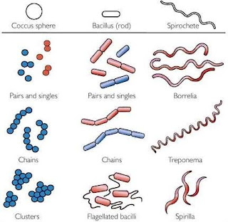
Fig. 2: Common Shapes of Bacteria
Source: Microorganisms in our World by Atlas (1995)
In addition to the common bacterial shapes, many others also occur in different shapes, which include:
- pear shaped cells, e.g. Pasteuri
- lobed spheres, e.g. Sulfolobus
- rods with squared ends, e.g. Bacillus anthracis
- disk arranged stacks of coins, e.g. Caryophanon
- rods with helically sculptured surfaces, e.g. Seliberia and many others.
Bacterial Structures
Examination of a bacterial cell will reveal several components and structures. Some are external to the cell wall while others are internal to the cell wall.Structure External to the Cell Wall
- Flagella (Singular: Flagellum): These are hair like, helical appendages that protrude through the cell wall, 0.01 – 0.02µm in diameter and simple in structure. Based on their location on the cell, flagella may be polar or lateral.
- Polar: At one or both ends of bacterium.
- Lateral: Along the sides of the bacterium.
- A based body associated with the cytoplasmic, membrane and cell wall.
- A short hook and a helical filament which is usually several times as long as the cell.
- A flagellum grows at the tip rather than at the base.
Types of Flagella
- Monotrichous: A single polar flagellum. Many that appears and functions as monopolar or bipolar flagella consist of bundles of 2 to 50 single units (polytrichous).
- Lophotrichous: A cluster of polar flagella.
- Amphitrichous: Flagella, either single or clusters at both cell poles.
- Peritrichous: Cell surrounded by lateral flagella.
Function of Flagella
Bacteria propel themselves by rotating their helical flagella.
Fig. 3: Types of Flagella
Source: bioweb.uwlax.edu
2. Pili (Singular: Pilus): They are also called fimbriae. They are hollow, non-helical filamentous appendages that are thinner, shorter and more numerous than flagella: long, thin, straight threads 3-25µm in diameter and 12µm in length. They do not function in motility since they are found on non-motile and motile species. Several functions are associated with different types of pili.
F pilus (Sex pilus) serves as the path of entry of genetic material during bacterial mating. Some play major role in human infection by allowing pathogenic bacteria to attach to the epithehal cells
lining the respiratory, intestinal or genitourinary tracts, this prevents the bacteria from being washed away by the flow of mucous or body fluids and permits infections to be established.
Social Plugin